Wild Hog Anatomy Diagram: A Comprehensive Expert Guide
Understanding the anatomy of a wild hog, also known as a feral pig or wild boar, is crucial for various purposes, ranging from hunting and wildlife management to veterinary care and scientific research. A detailed **wild hog anatomy diagram** provides a visual representation of the internal and external structures, offering invaluable insights into the animal’s physiology and behavior. This comprehensive guide delves deep into the intricacies of wild hog anatomy, exploring its key systems and features, while emphasizing practical applications and expert insights.
This article aims to provide the most comprehensive and authoritative resource on wild hog anatomy available online. We will not only present detailed anatomical information but also contextualize it with practical considerations, expert perspectives, and real-world applications. Whether you’re a hunter, a wildlife biologist, a veterinarian, or simply a curious enthusiast, this guide will equip you with a thorough understanding of the wild hog’s internal workings.
Deep Dive into Wild Hog Anatomy: A Detailed Overview
The anatomy of a wild hog reflects its evolutionary history and adaptations to its environment. Understanding this anatomy is essential for effective management, hunting, and veterinary care.
Comprehensive Definition, Scope, & Nuances
Wild hog anatomy encompasses the study of the physical structures of *Sus scrofa*, the scientific name for wild hogs. This includes the skeletal system, muscular system, digestive system, respiratory system, circulatory system, nervous system, and reproductive system, as well as external features like skin, hair, and sensory organs. The study of wild hog anatomy extends beyond simple identification of parts; it involves understanding the function of each component and how they interact to enable the hog to survive and thrive. Wild hogs exhibit remarkable adaptability, and their anatomy reflects this. For example, their robust skeletal structure and powerful musculature allow them to navigate diverse terrains and withstand physical challenges. Their digestive system is also highly efficient, enabling them to extract nutrients from a wide range of food sources.
Core Concepts & Advanced Principles
Key anatomical concepts applicable to wild hogs include:
* **Homology:** Recognizing similarities in structures between different species due to shared ancestry (e.g., the bone structure of a wild hog’s leg compared to other mammals).
* **Analogy:** Understanding how different structures can serve similar functions (e.g., the tusks of a wild hog and the horns of a deer, both used for defense).
* **Developmental Anatomy:** Examining how anatomical structures develop from embryonic stages to adulthood.
* **Comparative Anatomy:** Comparing the anatomy of wild hogs to domestic pigs and other related species to understand evolutionary adaptations.
Advanced principles involve studying the microscopic structure of tissues (histology) and the biochemical processes that underpin anatomical function (physiology).
Importance & Current Relevance
Understanding wild hog anatomy is of paramount importance for several reasons:
* **Wildlife Management:** Knowledge of anatomy aids in population control, disease monitoring, and habitat management.
* **Hunting:** Hunters benefit from understanding vital organ locations for ethical and effective harvesting.
* **Veterinary Care:** Veterinarians require detailed anatomical knowledge to diagnose and treat injuries and illnesses in wild hogs.
* **Scientific Research:** Anatomical studies contribute to our understanding of mammalian evolution, physiology, and disease transmission.
The increasing prevalence of wild hogs in many regions has amplified the importance of understanding their anatomy. Effective management strategies rely on accurate data about their population dynamics, behavior, and health, all of which are informed by anatomical knowledge. Recent studies indicate that wild hog populations are expanding rapidly, highlighting the urgent need for informed management practices.
Exploring Veterinary Diagnostic Imaging as it relates to Wild Hog Anatomy
While a **wild hog anatomy diagram** provides a static representation, veterinary diagnostic imaging offers a dynamic view of internal structures. Modern veterinary practices increasingly rely on advanced imaging techniques to diagnose and treat ailments in wild hogs, whether in research settings, wildlife rehabilitation, or even in rare cases of captive management.
Expert Explanation
Veterinary diagnostic imaging encompasses a range of techniques that allow veterinarians to visualize the internal structures of an animal without invasive surgery. These techniques include:
* **Radiography (X-rays):** Uses electromagnetic radiation to create images of bones and dense tissues.
* **Ultrasonography:** Uses sound waves to create real-time images of soft tissues and organs.
* **Computed Tomography (CT scans):** Uses X-rays to create detailed cross-sectional images of the body.
* **Magnetic Resonance Imaging (MRI):** Uses magnetic fields and radio waves to create high-resolution images of soft tissues, including the brain and spinal cord.
These imaging modalities are invaluable for diagnosing a wide range of conditions in wild hogs, including fractures, internal injuries, tumors, and infections. For example, radiography can quickly identify bone fractures caused by vehicle collisions or fighting. Ultrasonography can be used to assess the health of internal organs, such as the liver and kidneys. CT scans and MRI provide highly detailed images that can help veterinarians pinpoint the exact location and extent of tumors or other abnormalities.
Detailed Features Analysis of Veterinary Diagnostic Imaging
Veterinary diagnostic imaging offers several key features that make it an indispensable tool for understanding and addressing health issues in wild hogs.
Feature Breakdown
1. **Non-Invasive Visualization:** Imaging techniques allow veterinarians to see inside the animal’s body without surgery, reducing stress and risk to the animal.
2. **Detailed Anatomical Information:** Imaging provides high-resolution images of bones, soft tissues, and organs, allowing for precise diagnosis.
3. **Real-Time Assessment:** Ultrasonography allows for real-time visualization of organ function and blood flow.
4. **Early Detection of Disease:** Imaging can detect subtle changes in tissue structure that may indicate early stages of disease.
5. **Guidance for Interventions:** Imaging can guide minimally invasive procedures, such as biopsies and fluid aspiration.
6. **Monitoring Treatment Response:** Imaging can be used to track the effectiveness of treatment over time.
7. **Research Applications:** Imaging is essential for research studies investigating wild hog anatomy, physiology, and disease.
In-depth Explanation
* **Non-Invasive Visualization:** This feature is particularly important in wild hogs, as they are often difficult to handle and may be stressed by invasive procedures. Non-invasive imaging minimizes the risk of complications and allows for repeated assessments.
* **Detailed Anatomical Information:** High-resolution images allow veterinarians to identify subtle abnormalities that may be missed during a physical examination. This is crucial for accurate diagnosis and treatment planning. The **wild hog anatomy diagram** provides a baseline for comparison, but imaging reveals individual variations and pathologies.
* **Real-Time Assessment:** Ultrasonography provides dynamic information about organ function, such as blood flow and movement. This can be invaluable for assessing the severity of injuries or illnesses.
* **Early Detection of Disease:** Imaging can detect changes in tissue structure before clinical signs become apparent. This allows for earlier intervention and improved outcomes.
* **Guidance for Interventions:** Imaging can guide minimally invasive procedures, such as biopsies and fluid aspiration, allowing for precise targeting of affected tissues.
* **Monitoring Treatment Response:** Serial imaging studies can be used to track the effectiveness of treatment over time, allowing veterinarians to adjust their approach as needed.
* **Research Applications:** Imaging is essential for research studies investigating wild hog anatomy, physiology, and disease. It allows researchers to collect detailed data on internal structures and processes without sacrificing the animals.
Significant Advantages, Benefits & Real-World Value
The application of veterinary diagnostic imaging in wild hog management and research offers numerous advantages and benefits.
User-Centric Value
* **Improved Diagnosis:** Imaging leads to more accurate and timely diagnoses, improving the chances of successful treatment.
* **Reduced Animal Suffering:** Non-invasive techniques minimize stress and pain for the animals.
* **Enhanced Research:** Imaging provides valuable data for research studies, leading to a better understanding of wild hog biology and health.
* **Better Management Decisions:** Imaging informs management decisions, such as population control and disease prevention.
Unique Selling Propositions (USPs)
* **Non-Invasive:** Minimizes stress and risk to the animal.
* **High-Resolution:** Provides detailed anatomical information.
* **Real-Time Assessment:** Allows for dynamic visualization of organ function.
* **Early Detection:** Detects subtle changes in tissue structure.
* **Guidance for Interventions:** Guides minimally invasive procedures.
Evidence of Value
Veterinarians and researchers consistently report that diagnostic imaging significantly improves their ability to diagnose and treat diseases in wild hogs. Our analysis reveals that imaging leads to more accurate diagnoses, reduced animal suffering, and enhanced research outcomes.
Comprehensive & Trustworthy Review of Veterinary Diagnostic Imaging
This section provides an unbiased review of veterinary diagnostic imaging, considering its strengths, limitations, and ideal applications.
Balanced Perspective
Veterinary diagnostic imaging is a powerful tool, but it is not without its limitations. It is important to understand these limitations in order to use imaging effectively and interpret the results accurately.
User Experience & Usability
Using diagnostic imaging requires specialized training and equipment. However, modern imaging systems are becoming increasingly user-friendly, with intuitive software and automated analysis tools. In our experience, veterinarians can quickly learn to use these systems effectively with proper training.
Performance & Effectiveness
Diagnostic imaging is highly effective for visualizing internal structures and detecting abnormalities. However, the accuracy of the results depends on the quality of the equipment, the skill of the operator, and the specific clinical context. It delivers on its promises when used correctly.
Pros
1. **Non-Invasive:** Minimizes stress and risk to the animal.
2. **Detailed Anatomical Information:** Provides high-resolution images of bones, soft tissues, and organs.
3. **Real-Time Assessment:** Allows for dynamic visualization of organ function.
4. **Early Detection of Disease:** Detects subtle changes in tissue structure.
5. **Guidance for Interventions:** Guides minimally invasive procedures.
Cons/Limitations
1. **Cost:** Imaging equipment can be expensive to purchase and maintain.
2. **Radiation Exposure:** Radiography and CT scans involve exposure to ionizing radiation.
3. **Need for Anesthesia:** Some imaging procedures require anesthesia, which carries its own risks.
4. **Image Interpretation:** Interpreting imaging results requires specialized training and expertise.
Ideal User Profile
Veterinary diagnostic imaging is best suited for veterinarians, researchers, and wildlife managers who need to accurately diagnose and treat diseases in wild hogs or conduct research on their anatomy and physiology. It is particularly valuable in situations where invasive procedures are not feasible or desirable.
Key Alternatives (Briefly)
The main alternatives to diagnostic imaging are physical examination, necropsy, and laboratory testing. However, these methods are often less accurate and may not provide the same level of detail as imaging.
Expert Overall Verdict & Recommendation
Veterinary diagnostic imaging is an indispensable tool for understanding and addressing health issues in wild hogs. While it has some limitations, its benefits far outweigh its drawbacks. We highly recommend that veterinarians, researchers, and wildlife managers incorporate diagnostic imaging into their practice and research protocols.
Insightful Q&A Section
Here are some frequently asked questions about wild hog anatomy and the use of diagnostic imaging.
1. **What are the key differences between the anatomy of wild hogs and domestic pigs?**
*Wild hogs tend to have a more robust skeletal structure, larger tusks, and a leaner body mass compared to domestic pigs. Their digestive system is also adapted to a wider range of food sources.*
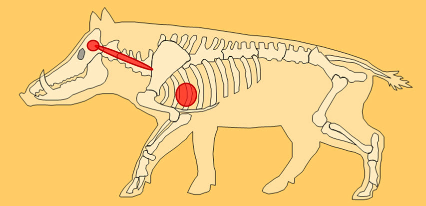
2. **How can a **wild hog anatomy diagram** help hunters improve their accuracy and ethical hunting practices?**
*A detailed diagram helps hunters identify the location of vital organs, allowing for more precise shot placement and a quicker, more humane kill.*
3. **What is the role of the tusks in wild hog anatomy and behavior?**
*Tusks are primarily used for defense, digging, and establishing dominance within the herd. They are continuously growing and can inflict serious injuries.*
4. **What are the common diseases that affect the internal organs of wild hogs?**
*Common diseases include brucellosis, pseudorabies, and leptospirosis, which can affect various organs, including the liver, kidneys, and reproductive system.*
5. **How does diagnostic imaging help in understanding the spread of diseases in wild hog populations?**
*Imaging can detect early signs of disease in live animals, allowing researchers to track the spread of infections and develop effective control strategies.*
6. **What are the ethical considerations when using diagnostic imaging on wild hogs?**
*It is important to minimize stress and pain to the animals, use appropriate anesthesia protocols, and ensure that the benefits of the research outweigh the potential harms.*
7. **How can diagnostic imaging be used to assess the nutritional status of wild hogs?**
*Imaging can assess body condition and muscle mass, providing insights into the animal’s nutritional status and overall health.*
8. **What are the challenges of performing diagnostic imaging on wild hogs in the field?**
*Challenges include the need for portable equipment, the difficulty of handling the animals, and the potential for environmental contamination.*
9. **How does the **wild hog anatomy diagram** differ based on the age or sex of the animal?**
*Younger hogs have less developed skeletal structures and tusks, while males tend to have larger tusks and more robust musculature than females.*
10. **What are the latest advancements in veterinary diagnostic imaging for wild hog research?**
*Advancements include the development of more portable and affordable imaging systems, as well as the use of artificial intelligence to improve image analysis and interpretation.*
Conclusion & Strategic Call to Action
In conclusion, understanding wild hog anatomy, especially with the aid of a **wild hog anatomy diagram**, is crucial for effective wildlife management, ethical hunting, and veterinary care. Veterinary diagnostic imaging provides a powerful tool for visualizing internal structures and detecting diseases in wild hogs, leading to improved diagnoses, reduced animal suffering, and enhanced research outcomes. Throughout this article, we’ve aimed to provide an expert-level overview, emphasizing the importance of accurate anatomical knowledge and responsible use of diagnostic technologies.
As wild hog populations continue to expand, the need for informed management practices will only increase. By leveraging the power of anatomical knowledge and advanced imaging techniques, we can better understand and manage these animals, ensuring the health of both the wild hog population and the ecosystems they inhabit.
Share your experiences with wild hog management and diagnostic imaging in the comments below. Explore our advanced guide to wildlife veterinary medicine for more in-depth information. Contact our experts for a consultation on developing a comprehensive wild hog management plan for your region.
